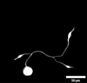
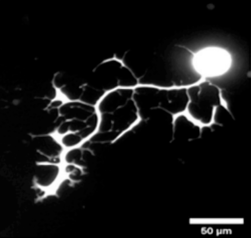
Original neuron images from: “Cho et al. Neurite growth of trigeminal ganglion neurons in vitro with near-infrared light irradiation Journal of Photochemistry and Photobiology B: Biology 210 111959”
https://doi.org/10.1016/j.jphotobiol.2020.111959
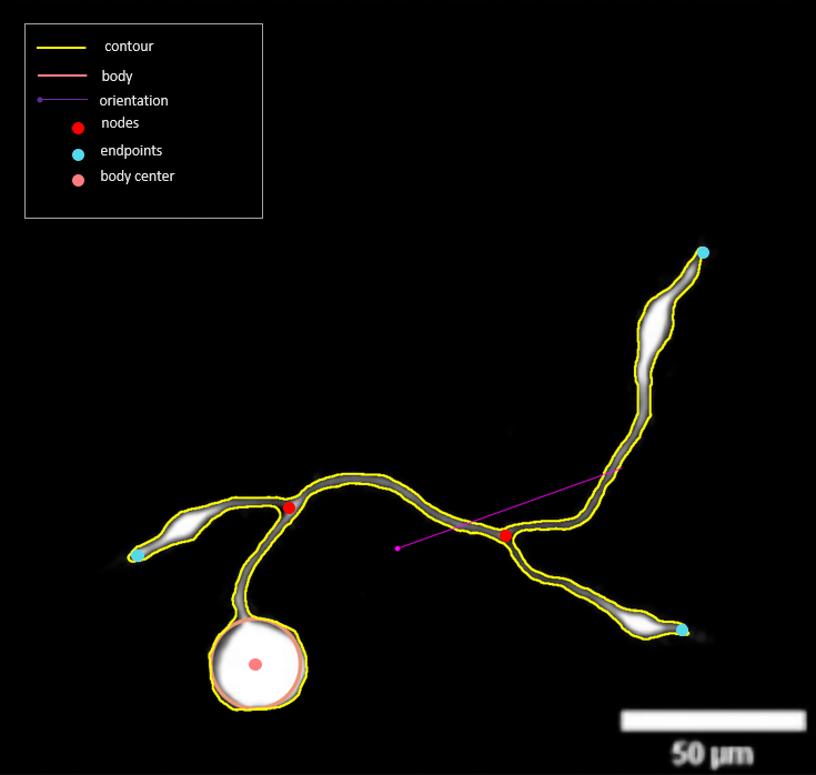
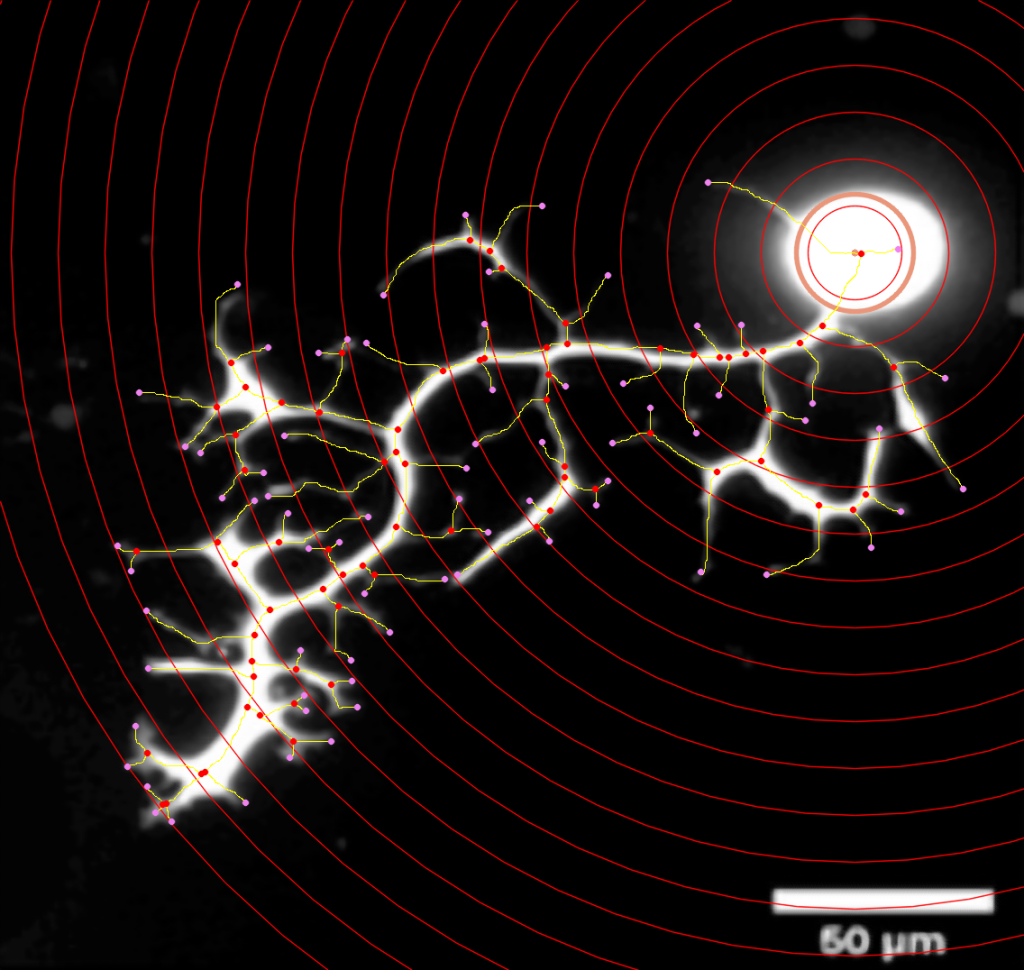
Images were analysed with the automatic Sholl analysis module in the KARMENscience. Automatic segmentation and classification of the neurons. Left image also shows the principal orientation of the neuron, together with the position of the center-of-gravity of the segmented neuron. The right image shows Sholl rings with endpoints in all branches.
*Disclaimer: This page is available only by a direct link to whoever has been contacted by KARMENscience team.